 Products Home
Veneto® Inverted Microscopes

Please Wait
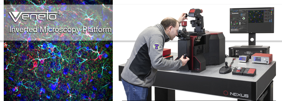
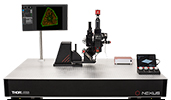
With widefield, confocal, and multiphoton imaging options, a built-in piezo focusing module, and an integrated breadboard to create your own optical paths, this microscope is ideal for today and for 10 years from now.

Sam Tesfai
General Manager,
Feedback?
Questions?
Need a Quote?


Veneto® Inverted Microscope Systems
Thorlabs' Modular Inverted Microscopy Platform provides a turnkey solution for widefield, confocal, and multiphoton imaging. Inverted microscopes are powerful and versatile research tools that can accommodate a range of applications, such as fluorescence, in vivo, ex vivo, 3D, high-resolution, high-speed (video-rate), and live tissue imaging. This platform is designed to meet the needs of labs working in cell biology and other life science applications that require an inverted microscopy platform while including key features provided by our upright systems, such as multiple imaging modalities, accessory ports, and easily accessible light paths.
The key highlights that set our Veneto inverted microscope platform apart from the typical inverted microscope are listed in the Highlights tab, while the Features tab gives more details on the supported imaging techniques and the day-to-day usage features. This platform is available as a turnkey system that also offers users access to internal optomechanics and optics for DIY customization. We offer configurations that support widefield, brightfield, phase, Dodt, confocal, and multiphoton imaging; see the Configurations tab for more details.
Options at a Glance
Laser Scanning
- 8 kHz and 12 kHz Resonant-Galvo-Galvo (US Patent 10,722,977) and Galvo-Resonant Scanners for High-Speed Imaging
- Galvo-Galvo Scanners for User-Defined ROI Shapes and Photostimulation Patterns
- Super Broadband Scan Optics Optimized for Two-Photon and Three-Photon Imaging
Signal Detection
- Standard Multialkali or High-Sensitivity GaAsP PMTs
- 13° Detection Module for up to Four PMTs with Confocal Imaging
- 13° Non-Descanned Detection Module for Two PMTs with Multiphoton Imaging
Up to 7 Customizable Optical Pathways
- 3 Paths on Bottom Tier for Laser Scanning and Widefield Imaging
- 1 Path on Second Tier for Reflected Light Illumination or Laser Scanning
- 1 Path on Third Tier Behind the Objective for Non-Descanned Detection
- 1 Transmitted Light Path
- 1 Trinocular Path
If you have additional questions about our Veneto imaging systems, please click on the Contact Me button to send us an email.
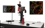
Credit for Image Above: Wild-type mouse brain section (300 μm x 300 μm) tagged with DAPI (405 nm), Alexa 488 anti-S100B, Alexa 555 anti-Neurofilament, and Alexa 633 anti-GFAP. Sample Courtesy of Lynne Holtzclaw, NIH/NICHD/MIC.
| Table 2.1 Quick Links |
|---|
| Vertically Integrated System |
| High-Speed Scanners |
| Built-In Motorized Focusing Module |
Veneto® Highlights
The Veneto Inverted Microscopes are turnkey systems that support widefield, confocal, and multiphoton imaging. This tab outlines the key highlights that set the Veneto platform apart from the typical inverted microscope. See Table 2.1 for the quick links to each section.
Click on different parts of the microscope for more details.
Vertically Integrated System
- Composed of Thorlabs Manufactured Components
- Multiple Ports for Mounting Scientific Cameras or Detection Devices
- Bottom Port for Efficient Emission Detection (Requires Custom Optical Table with Compatible Opening for Access; To Request More Information, Click Here)
- Motorized Six-Position Turret Holds Ø32 mm Excitation and Emission Filters to Support Greater Collection Angles for Increased Signal
- High-Speed Motorized XY Stage with Scanning Speed up to 250 mm/s
- Light Sources
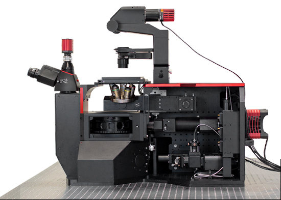
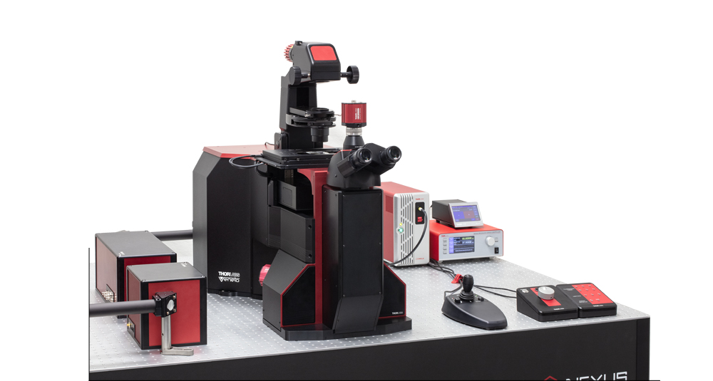
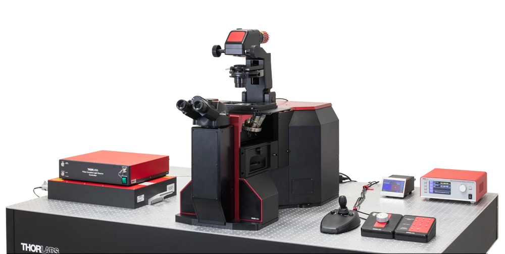
Our Veneto microscopes come fully integrated with all the necessary components required to meet the exact needs of an experiment. As shown in Figure 2.2, Thorlabs manufactures the components to ensure seamless functionality; click on different parts of the microscope for more details. With a compact 32" x 18" (812.8 mm x 457.2 mm) footprint, our inverted microscopes offer broad compatibility with a variety of light sources and accept Ø32 mm optics in a motorized six-position filter turret. Direct access to several independent optical paths via SM2 (2.035"-40) ports with 60 mm cage pattern holes or SM30 (M30.5 x 0.5) ports allows researchers the option to build in their own custom imaging modalities.
Integrated Control Unit and ThorImage®LS Software
The included ThorImageLS software supports multiple imaging techniques and offers seamless modality changes when switching between configurations. An electronic control unit allows the user to operate all included motorized components through ThorImageLS and handheld controllers. The software recognizes each component once plugged in. Additionally, the control unit offers empty slots for future upgradability to add more modalities. Users can customize their workspace in the intuitive GUI and choose to visualize only the settings relevant to each aspect of the experiment. Open SDKs enable users to code their own device plugins; see the ThorImageLS tab for more details.
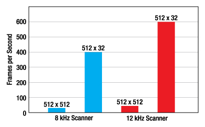
Figure 2.3 Galvo-Resonant Scanners offer high frame rates for high-speed imaging. The 8 kHz scanners offers a maximum frame rate up to 400 fps for 512 x 32 resolution. The 12 kHz scanners offer a maximum frame rate up to 600 fps for a 512 x 32 resolution.
High-Speed Scanners
- 8 kHz and 12 kHz Galvo-Resonant Scanners for High-Speed Imaging
- Galvo-Galvo Scanners for User-Defined ROI Shapes and Photostimulation Patterns
- 8 kHz and 12 kHz Resonant-Galvo-Galvo (US Patent 10,722,977) for Sequential Targeting
Veneto inverted microscopes can be configured with one or two co-registered scan paths to propagate, condition, and direct an input laser beam. We offer galvo-resonant scanners, a galvo-galvo scanner, and resonant-galvo-galvo scanners. These choices allow the user to optimize each experiment as needed for high frame rates, high sensitivity, and/or targeted exposure of the regions of interest.
Galvo-Resonant Scanners for High-Speed Imaging: Thorlabs offers 8 kHz and 12 kHz galvo-resonant scanners. Our 8 kHz scanners utilize the entire field of view and offer a maximum frame rate of 400 fps, while our 12 kHz scanners provide an increased frame rate of 600 fps.
Galvo-Galvo Scanners for User-Defined ROI Shapes: Galvo-galvo scanners support user-drawn scan geometries (lines, polylines, squares, and rectangles) and also support custom photoactivation patterns (circles, ellipses, polygons, and points). They offer consistent pixel dwell times for better signal integration and image uniformity.
Resonant-Galvo-Galvo Scanners for Multimodal Scanning: Thorlabs offers 8 kHz and 12 kHz resonant-galvo-galvo (RGG) scanners. The design of our RGG scanners is registered under US Patent 10,722,977. These scanners use an additional galvanometer in concert with a traditional galvo-resonant scanner to move the scan region. Edges of the scan region and the path the beam takes between scan regions are masked by Pockels cells to avoid unwanted exposure. Our 8 kHz scanners utilize the entire field of view and offer a maximum frame rate of 400 fps, while our 12 kHz scanners provide an increased frame rate of 600 fps.

Click to Enlarge
Figure 2.4 The built-in motorized focusing module translates the entire third tier pathway (outlined in red). This pathway includes the objective turret and the non-descanned PMTs.
Built-In Motorized Focusing Module
- Innovative Motorized Focusing Module Designed into the Microscope Frame
- Z-Stacks Can be Taken with Different Objectives by Turning the Objective Nosepiece
- Move Objectives Using Stepper Motor with 15 mm Travel Range and 50 nm Resolution
- Optional Piezo Motor with 100 µm Travel Range and 50 nm Resolution
- Control Focusing Module via the Included ThorImageLS Software
The Veneto microscope platform features an innovative built-in motorized focusing module fitted with a absolute linear encoder and a compact stepper motor actuator. The actuator provides smooth linear motion control and allows very small step sizes over the entire 15 mm travel range, delivering greater flexibility with a fine 50 nm resolution. The focusing module translates the entire third tier pathway that lies behind the objective; see Figure 2.4. This means a Z-stack can be easily taken with different objectives by simply turning the nosepiece to a new objective. This pathway is mounted on a vertical stage, which has a rigid, all-steel design and heavy-duty cross-roller bearings for uniform performance over the entire range of motion. The focusing module, which is built into the microscope frame behind the pathway, drives the stage to translate the objective turret and non-descanned PMTs.
For more precise movement, the focusing module can include an optional piezo motor that offers 100 µm travel with 50 nm resolution. The dual-drive option for the focusing module allows users to image multiple z-stacks by using the stepper motor and piezo simultaneously. Users can control the focusing module via the included ThorImageLS software.
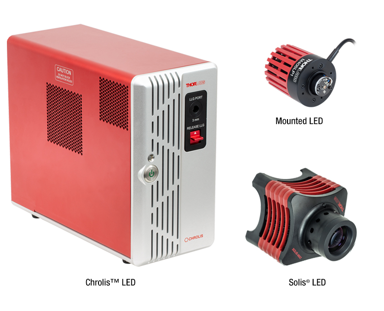
Mounted LEDs
- Wavelengths Ranging from 265 nm to 1650 nm
- White, Broadband, and Dual-Peak LEDs Also Available
- Thermal Properties Optimized for Stable Output Power
Solis®High-Power Mounted LEDs
- Typical Collimated Output Powers from 570 mW to 7.1 W
- Fanless Design Efficiently Dissipates Heat without Introducing Vibrations
- 20 Wavelengths Available
Chrolis™ 6-Wavelength High-Power LED Sources
- Combine 6 User-Changeable, High-Power LEDs into One Liquid Light Guide
- Eleven Wavelength Options Cover All Known Fluorophores Between 365 nm and 780 nm
- Pre-Configured or Custom Configurations Available
Click to Enlarge
Illumination Sources
- Ultra Fast XY Scanning Up to 250 mm/s
- Integrated Brushless DC Linear Servo Motor Actuators
- 110 mm x 75 mm (4.3" x 2.95") Travel Range
- High Repeatability (0.25 µm) and Position Accuracy (<3 µm)
- See the Complete Website Presentation
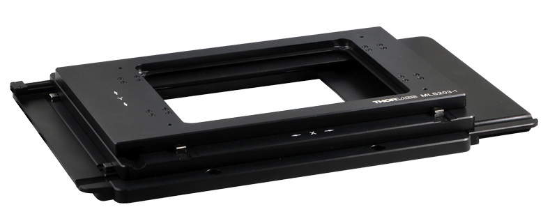
Click to Enlarge
High-Speed Motorized XY Scanning Stage
- Zelux™ Monochrome or Color CMOS Cameras for General-Purpose Imaging Applications
- Kiralux® CMOS Cameras for a Range of Applications, Including Fluorescence Microscopy, Materials Inspection, and Multispectral Imaging
- Monochrome, Color, NIR-Enhanced, or Polarization-Sensitive Sensors Available
- Quantalux® Monochrome sCMOS Cameras for Low-Light Applications
- Version Available with Hermetically Sealed TE-Cooled Packaging
- See Our Complete Range of Scientific Cameras.
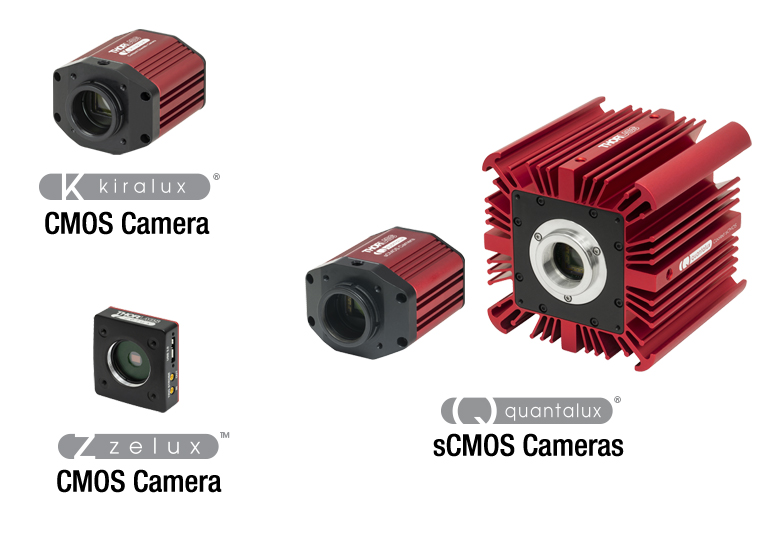
Click to Enlarge
Camera Housings of Our Compact Scientific and Scientific CCD Cameras
MCWHLP1 Mounted LED
- 6500 K (Cold White) Color Temperature
- 2350 mW (Min) Output Power
- 700 mA Max Current (CW)
- Typical Lifetime >100,000 Hours
- Ideal for Fluorescence Microscopy Applications
This is an excerpt from the 2021 Focus on Microscopy Meeting with Jeff Brooker, our CTO for Life Sciences.
Veneto® Features
The Veneto Inverted Microscopes are turnkey systems that support widefield, confocal, and multiphoton imaging. This tab gives details on the different types of imaging techniques, beam conditioning modules, included software, and Thorlabs support. For a walkthrough on the different modules available with the Veneto Inverted Microscope, see Video 3.1.
| Imaging Techniques | ||
|---|---|---|
| Scan Paths Designed In-House |
|
|
| Scanner Options | Resonant- Galvo-Galvo |
|
| Galvo-Resonant |
|
|
| Galvo-Galvo |
|
|
| Confocal Imaging |
|
|
| Multiphoton Imaging |
|
|
| Widefield Viewing |
|
|
| Reflected Light Illumination | ||
| Transmitted Light Imaging |
|
|
| Beam Conditioning Modules for Multiphoton Imaging | ||
|---|---|---|
| Fast Laser Power Modulators |
|
|
| Pockels Cells |
|
|
| Variable Attenuator |
|
|
| Beam Stabilizer |
|
|
| Day-to-Day Usage | ||
|---|---|---|
| Several Software Packages |
|
|
| Input and Output Triggers |
|
|
| Thorlabs Support | ||
|---|---|---|
| Fully Designed and Manufactured In-House |
|
|
| Professional Installation |
|
|
| Quick Support |
|
|
Thorlabs recognizes that each imaging application has unique requirements.
If you have any feedback, questions, or need a quotation, please use our
inverted microscope contact form or call (703) 651-1700.
Example Configurations
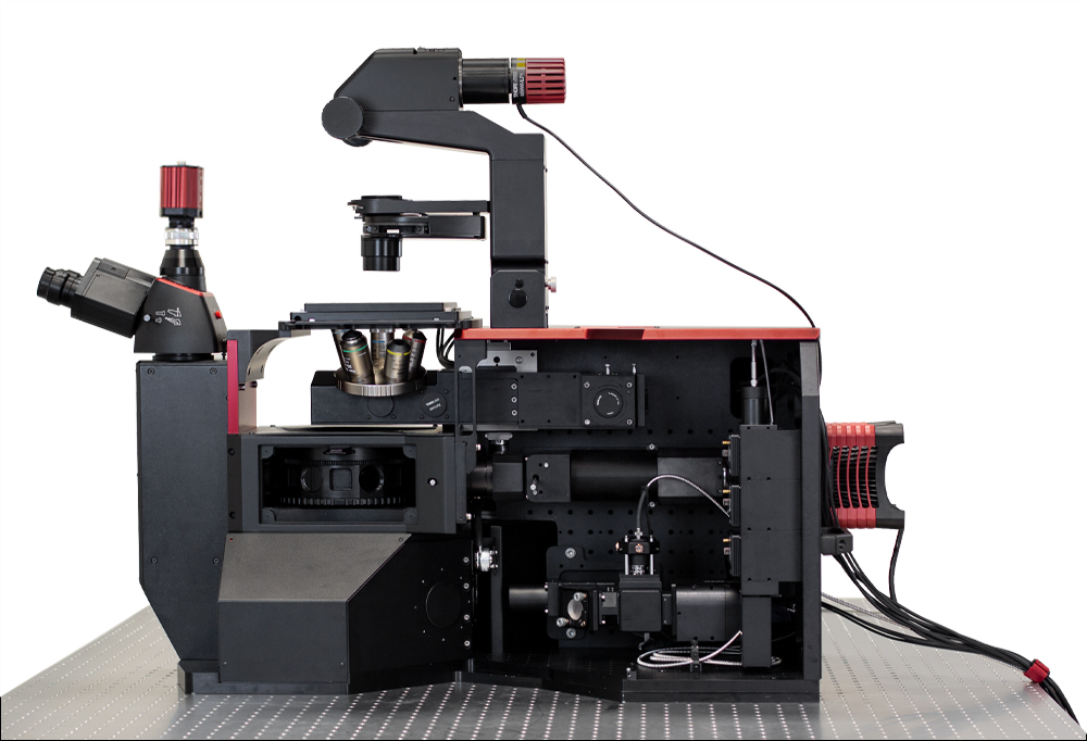
The Veneto® Inverted Microscopy Platform is designed to enable application-specific configurations. We currently offer confocal and two-photon configurations; two example configurations are shown here. Both configurations additionally support widefield viewing. As shown in Figures 4.1 and 4.2, users can remove the side cover to access several independent optical pathways. DIY customization is possible via SM2 (2.035"-40) ports with 60 mm cage pattern holes or SM30 (M30.5 x 0.5) ports if users want to build in their own custom imaging modalities.
Two-Photon + Widefield ConfigurationFeatures
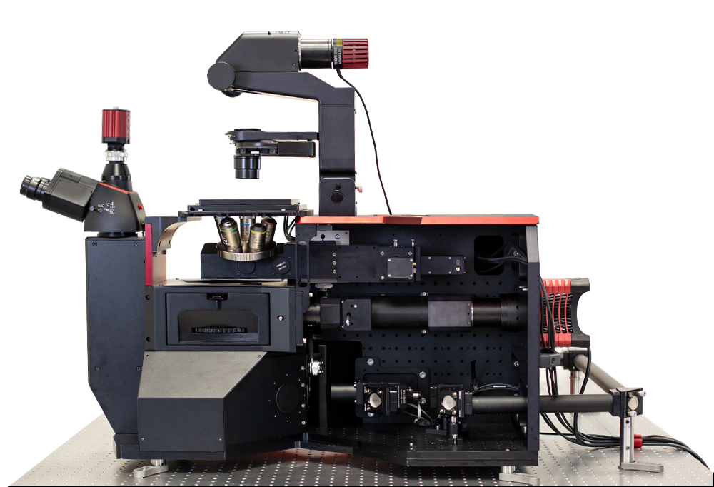 Click to Enlarge Figure 4.1 Two-Photon + Widefield Configuration with the Side Cover Removed |
Confocal + Widefield ConfigurationFeatures
 Click to Enlarge Figure 4.2 Confocal + Widefield Configuration with the Side Cover Removed |
![]()
The full source code for ThorImage®LS is available for owners of a Bergamo®, Veneto®, or Confocal microscope. Click here to receive your copy.

ThorImage®LS Software
ThorImageLS is an open-source image acquisition program that controls Thorlabs' microscopes, as well as supplementary external hardware. From prepared-slice multiphoton Z-stacks to simultaneous in vivo photoactivation and imaging, ThorImageLS provides an integrated, modular workspace tailored to the individual needs of the scientist. Its workflow-oriented interface supports single image, Z-stacks, time series, and image streaming acquisition, visualization, and analysis. See Video 121A for a real-time view of data acquisition and analysis with ThorImageLS.
ThorImageLS is included with a Thorlabs microscope system purchase and is open source, allowing full customization of software features and performance. ThorImageLS also includes Thorlabs’ customer support and regular software updates to continually meet the imaging demands of the scientific community.
For additional details, see the full web presentation.
Advanced Software Functionality
- Multi-Column Customizable Workspace
- Image Acquisition Synced with Hardware Inputs and Timing Events
- Live Image Correction and ROI Analysis
- Independent Galvo-Galvo and Galvo-Resonant Scan Areas and Geometries
- Tiling for High-Resolution Large-Area Imaging
- Independent Primary and Secondary Z-Axis Control for Fast Deep-Tissue Scans
- Automated Image Capture with Scripts
- Compatible with ImageJ Macros
- Multi-User Settings Saved for Shared Workstations
- Individual Colors for Detection Channels Enable Simple Visual Analysis
Seamless Integration with Experiments
- Simultaneous Multi-Point Photoactivation and Imaging with Spatial Light Modulator
- Fast Z Volume Acquisition with PFM450E or Third-Party Objective Scanners
- Electrophysiology Signaling
- Wavelength Switching with Coherent Chameleon Lasers
- Pockels Cell ROI Masking
- Power Ramped with Depth to Minimize Damage and Maximize Signal-to-Noise
New Functionality: Version 4.3 (Click to Expand for More Details)
Please contact ImagingTechSupport@thorlabs.com to obtain the latest ThorImageLS version compatible with your microscope. Because ThorImageLS 4.x adds significant new features over 3.x, 2.x and 1.x versions, it may not be compatible with older microscopes. We continue to support older software versions for customers with older hardware. See the full web presentation for functionality of previous versions.
- Added Support for the Toptica iChrome CLE-50 Laser
- Added Support for 3P Imaging
- Added Support for the CS126MU, CS165MU, and CC505MU Monochrome Cameras
- Added Support for a Mini-Circuits® Switch Box
- Added Support for PMT3100R (Included in Some Bergamo Multiphoton Imaging Systems)
- Added Configurable Channel View Layout (Horizontal and Vertical)
- Added Improved Scan Path Realignment and Added Ability to Save Multiple Reference Images for Multiple Targets
- Allows for Simplified Relocation to Same Target Day to Day
- Added New Features for SLM Operation
- Added 3D Mode Toggle
- Added Z Offset
- Added Ability to Export Patterns
- Added Set Zero% and Delete All Buttons
- Added Pattern Center to Display as "0" Point
- Added SLM Control Panel Advanced Mode Enhancement
- Added New Optional Delay Between Epochs
- Added SLM Control Panel Import/Export Enhancement
- Ability to Export Table of SLM Patterns
- Added New SLM Settings
- Added to ThorSLMSettings.xml and Application Settings
- Added Ability to Offset the Z Position of Pattern Points in New 3D Mode
- Enhanced ORCA Fusion Features
- Updated Exposure Calculation for Master Pulse Mode
- Added Option to Enable Water Cooling Control
- Added Improved Performance When Switching Modalities
- Enhanced Two-Way Scanning
- Two-Way Scanning is Now Allowed Up to a Pixel Density of 4096 x 4096 When Only One Channel is Selected
- Only Selectable by Dragging the Slider Bar to the Desired Pixel Density in One-Way Before Switching to Two-Way
- Added Camera Frame Rate Control
- Added UI Control of Two Blue Mini-Circuits® Switch Boxes
- Added 3D SLM
- Added Continuous Preview and Enhanced Orthogonal Views for Z-Stacks
- Added Improved Laser Safety Control for Digital Switches When Switches Are Configured for Laser Switching
- Added Control for Stimulus Shutter Operation When Using Stimulus Capture Mode
- Added Camera Frame Rate Control
- Added a New Section to Control the Frame Rate for CMOS Cameras with this Functionality
- Added Image-Based Autofocus
- Added Ability to Find the Optimal Focus Point of the Sample Based on Image Contrast
- Added Automatic Version Update Checker
- When Connected to Internet a Version Update Check Will Occur as the Splash Screen Loads When ThorImageLS Is Started Up
- Added Signal Generator Analog Mode
- Allows Custom Control of Analog Modulation
- Added New Continuous Button for Repeated Preview
- Allows for Fast Location Sample in XZ and YZ Line Scan Mode
- Added Enhanced IPC Communication
- Added IPC Command to Load an Experiment or a Template
- Added IPC Commands to Move X, Y, Z, and Secondary Z Stages
- Added IPC Commands that Get Sent from ThorImageLS Every Time a File Is Saved During T Series Experiments
Video 121A Features of ThorImage®LS
| Supported Imaging Platforms | ||
|---|---|---|
 Veneto® Inverted Microscopes |
 Bergamo® III Multiphoton Microscopes |
 Confocal Imaging Systems |
| Veneto® Microscope Specifications | ||
|---|---|---|
| Optical System | Infinity Corrected | |
| Optical Field Number | 20 | |
| Laser Scanning | Scan Path Wavelength Range | 450 - 1100 nm, 680 - 1300 nm, or 800 - 1800 nm |
| Scan Paths | Resonant-Galvo-Galvo Scanner, Galvo-Resonant Scanners, or Galvo-Galvo Scanners; Single or Dual Scan Paths |
|
| 8 kHz Resonant-Galvo-Galvo or Galvo-Resonant Scan Speed |
2 fps at 4096 x 4096 Pixels 30 fps at 512 x 512 Pixels 400 fps at 512 x 32 Pixels |
|
| 12 kHz Resonant-Galvo-Galvo or Galvo-Resonant Scan Speed |
4.4 fps at 2048 x 2048 Pixels 45 fps at 512 x 512 Pixels 600 fps at 512 x 32 Pixels |
|
| Galvo-Galvo Scan Speed | 3 fps at 512 x 512 Pixels 48 fps at 512 x 32 Pixels 70 fps at 32 x 32 Pixels Pixel Dwell Time: 0.4 to 20 µs |
|
| Galvo-Galvo Scan Modes | Imaging: Line, Polyline, Square, or Rectangle Non-Imaging: Circle, Ellipse, Polygon, or Point |
|
| Field of View | 20 mm Diagonal Square (Max) at the Intermediate Image Plane [12 mm Diagonal Square (Max) for 12 kHz Scanner] |
|
| Scan Zoom | 1X to 16X (Continuously Variable) | |
| Scan Resolution | Up to 2048 x 2048 Pixels (Bi-Directional) [Up to 1168 x 1168 Pixels for 12 kHz Scanners] Up to 4096 x 4096 Pixels (Unidirectional) [Up to 2336 x 2336 Pixels for 12 kHz Scanners] |
|
| Observation Tube | Trinoculars with 10X Eyepieces and Camera Port for 1X Camera Tube with External C-Mount Threads |
|
| Brightfield Light Source | Mounted LED (6500 K Correlated Color Temperature, 2350 mW Min. Output Power) |
|
| Widefield Light Source | Thorlabs' Mounted, Solis®, and Chrolis™ LEDs or Optional Port for Industry-Standard Liquid Light Guides |
|
| Laser Scanning Light Source | Confocal Source Customizable with Up to 4 Visible Laser Lines Multiphoton Sources for Two-Photon and Three-Photon Imaging |
|
| Condenser | LWD 0.52 NA 3.5 mm Translation with Trans-Illumination Module |
|
| Motorized XY Scanning Stage | Travel Range | 110 mm x 75 mm (4.3" x 2.95") |
| Velocity (Max) | 250 mm/s | |
| Acceleration (Max) | 2000 mm/s2 | |
| Horizontal Load Capacity (Max) | 1.0 kg (2.2 lb) | |
| Objective Turret | Five Manual Positions with M25 x 0.75 Threads | |
| Focusing Unit | 15 mm of Motorized Travel Focus and Optional Autofocus Stepper Motor: 15 mm Travel Range, 50 nm Resolution Piezo: 100 µm Travel Range, 50 nm Resolution |
|
| Filter Turret | Six Motorized Positions for Ø32 mm Excitation and Emission Filters | |
| Control Units | Stage Joystick and Microscope Control Pad | |
Thorlabs recognizes that each imaging application has unique requirements.
If you have any feedback, questions, or need a quotation, please use our
inverted microscope contact form or call (703) 651-1700.

Click to Enlarge
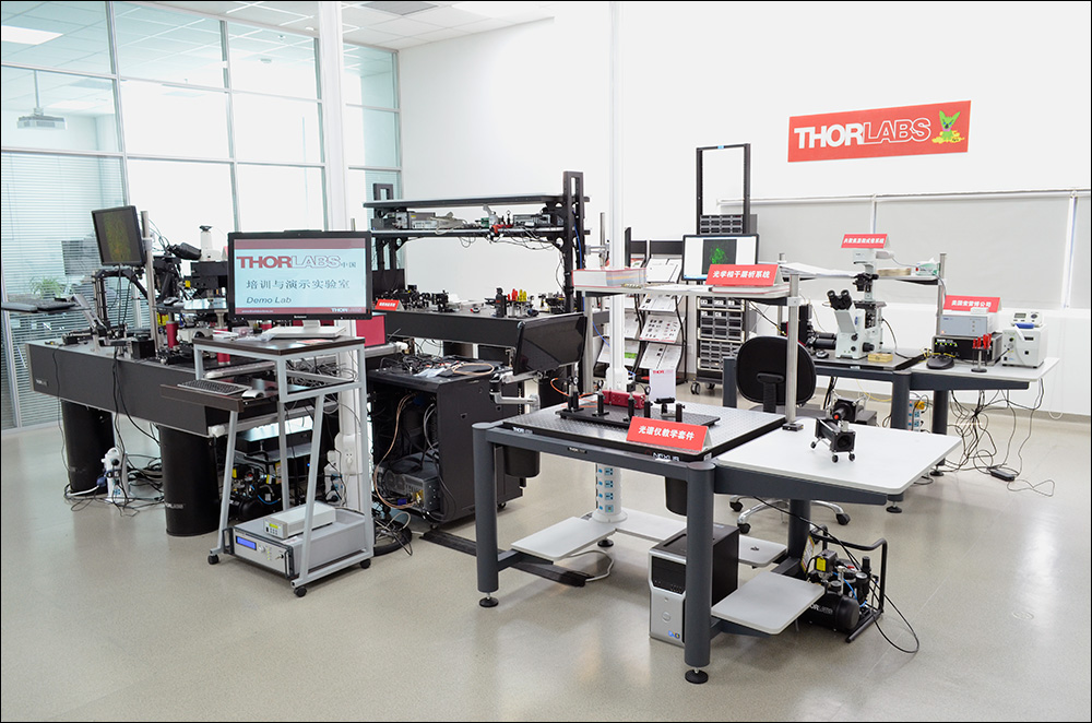
Figure 39A China Demo Room
Try Our Microscopes In Person or Virtually
Thorlabs' sales engineers and field service staff are based out of nine offices across four continents. We look forward to helping you determine the best imaging system to meet your specific experimental needs. Our customers are attempting to solve biology's most important problems; these endeavors require matching systems that drive industry standards for ease of use, reliability, and raw capability.
Thorlabs' worldwide network allows us to operate demo rooms in a number of locations where you can see our systems in action. We welcome the opportunity to work with you in person or virtually. A demo can be scheduled at any of our showrooms or virtually by contacting ImagingSales@thorlabs.com.
Customer Support Sites
(Click Each Location for More Details)
Newton, New Jersey, USA
Thorlabs Headquarters
43 Sparta Avenue
Newton, NJ 07860
Customer Support
- Phone: (973) 300-3000
- E-mail: techsupport@thorlabs.com
Ely, United Kingdom
Thorlabs Ltd.
1 Saint Thomas Place, Ely
Ely CB7 4EX
Customer Support
- Phone: +44 (0)1353-654440
- E-mail: techsupport.uk@thorlabs.com
Bergkirchen, Germany
Thorlabs GmbH
Münchner Weg 1
85232 Bergkirchen
Customer Support
- Phone: +49 (0) 8131-5956-0
- E-mail: europe@thorlabs.com
Puteaux, France
Thorlabs SAS
76 Route de la Demi Lune
92800 Puteaux
Customer Support
- Phone: +33 (0) 970 440 844
- E-mail: techsupport.fr@thorlabs.com
São Carlos, SP, Brazil
Thorlabs Vendas de Fotônicos Ltda.
Rua Rosalino Bellini, 175
Jardim Santa Paula
São Carlos, SP, 13564-050
Customer Support
- Phone: +55 (21) 2018 6490
- E-mail: brasil@thorlabs.com
Demo Rooms and Customer Support Sites
(Click Each Location for More Details)
Sterling, Virginia, USA
Thorlabs Imaging Systems HQ
108 Powers Court
Sterling, VA 20166
Customer Support
- Phone: (703) 651-1700
- E-mail: ImagingTechSupport@thorlabs.com
Demo Rooms
- Bergamo® Series Multiphoton Microscopes
- Veneto® Inverted Microscopes
- Upright Confocal Microscopy Systems
- Cerna Birefringence Imaging Microscopes
- Multiphoton Mesoscope
- OCT Systems: Telesto® and Ganymede®
Lübeck, Germany
Thorlabs GmbH
Maria-Goeppert-Straße 9
23562 Lübeck
Customer Support
- Phone: +49 (0) 8131-5956-40840
- Email: oct@thorlabs.com
Demo Rooms
- Ganymede® Series SD-OCT Systems
- Telesto® Series SD-OCT Systems
- Telesto® Series PS-OCT Systems
- Atria® Series SS-OCT Systems
- Vega™ Series SS-OCT Systems
Nerima-ku, Tokyo, Japan
Thorlabs Japan, Inc.
3-6-3 Kitamachi
Nerima-ku, Tokyo 179-0081
Customer Support
- Phone: +81-3-6915-7701
- Email: sales@thorlabs.jp
Demo Rooms
Shanghai, China
Thorlabs China
Room A101, No. 100, Lane 2891, South Qilianshan Road
Shanghai 200331
Customer Support
- Phone: +86 (0)21-60561122
- Email: techsupport-cn@thorlabs.com
Demo Rooms
- Bergamo® Series Multiphoton Microscopes
- Cerna Birefringence Imaging Microscopes
- OCT Systems: Telesto® and Ganymede®
| Posted Comments: | |
| No Comments Posted |